| Laboratory: | Oak Ridge National Laboratory (ORNL) |
| Capability Expert: | Karren L. More |
| Capability Details: | |
| Title: | Scanning transmission electron microscopy (STEM)-based 3D electron tomography |
| Class: | Characterization |
| Description: | STEM-based electron tomography is used to provide three-dimensional (3D) reconstructions of the structure (STEM image reconstruction) and composition (chemical map reconstruction) of materials. Electron tomography is analogous to bulk tomography techniques like X-ray tomography, but can provide information at a much higher resolution (sub-nm-scale) and can be used to combine morphological, crystallographic, and compositional data into 3D renderings. Electron tomography data can be acquired by using specialized microscopy holders, high-tilt tomography and cryo-tomography holders (tilt series through ±70°) and/or a 360° micropillar rotation holder, or through serial microtome sectioning. 3D electron tomography has many advantages over conventional 2D imaging and spectroscopic techniques and is a technique that can be used to interrogate complex structures, such as those comprised of multiple materials components, those with hierarchical porosity, structures with novel morphologies/geometries, and those that incorporate ultra-thin layers or coatings. |
| Capability Bounds: | NA |
| Unique Aspects: |
A full suite of scanning transmission electron microscopes (STEM) and unique tomography holders (high-angle tomography tilting, 360° rotation, and cryo-tilting tomography) are available at ORNL for acquiring 3D images and spectral data of PGM-free catalysts and membrane electrode assemblies (MEAs) from the bulk- to the nm-scale. Unique microscopes include a high-voltage (200kV) non-aberration-corrected STEM (FEI Talos) equipped with a high-collection-efficiency energy dispersive X-ray spectrometer (EDS) system, and an aberration-corrected STEM (300kV FEI Titan) equipped with both EDS and electron energy loss spectrometer (EELS), which enable the extremely rapid, automated, and efficient collection of structural, chemical, and compositional data from beam-sensitive PGM-free catalysts and MEA components (e.g., ionomer mapping and carbon-based materials) such that correlative 3D reconstructions of structure and composition can be produced. A full suite of 3D data acquisition control and reconstruction software is also available. STEM-based 3D electron tomography requires reproducible methods for preparing low-loadings of highly dispersed, clean powder specimens on thin support grids, and serial sectioning of specimens (MEAs, for example) through continuous slicing via ultramicrotomy. ORNL’s microscopy laboratories are fully equipped with state of the art sample preparation instruments, including a new Leica Ultramicrotome for diamond knife sectioning of intact MEAs. |
| Availability: | Instruments in ORNL’s Materials Characterization Center (MCC) are available for partnership with industry through Strategic Partnership Projects (SPP), cooperative research and development agreements (CRADAs) via full-cost recovery (hourly fee), and direct project collaborations to facilitate new materials discovery and understanding. Instruments in ORNL’s Center for Nanophase Materials Sciences (CNMS – a U.S. DOE Office of Science User Facility) are accessible through a peer-reviewed proposal process (no cost if results are publishable and full-cost recovery if data is proprietary). All instruments in the MCC and CNMS are available for characterization of PGM-free catalysts and MEAs. |
| References: |
H. Uchida, et al., “Electron Tomography of Nafion Ionomer Coated on Pt/Carbon Black in High Utilization Electrode for PEFCs,” Journal of Physical Chemistry B 110[27] 13319 (2006). T. Ito, et al., “Three-Dimensional Spatial Distributions of Pt Catalyst Nanoparticles on Carbon Substrates in Polymer Electrolyte Fuel Cells,” Electrochemistry 79[5] 374 (2011). |
| Benefit: | STEM-based 3D electron tomography can provide unique insight regarding PGM-free catalyst particle morphologies, hierarchical porosity, and MEA structures (including ionomer dispersions) and corresponding measurements of size and constituent distributions. |
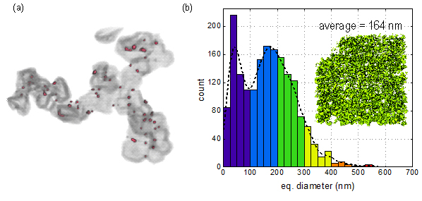
(a) 3D rendering of Pt nanoparticles supported on graphitized carbon. (b) 3D rendering of ionomer distribution within catalyst layer and size distribution of ionomer aggregates.